Kare Skin Cancer Center · Kare Plastic Surgery · Santa Monica
Melanoma Skin Cancer Center Santa Monica
804 7th Street, Santa Monica, CA 90403 · (310) 998-5533
Dermoscopy-enhanced melanoma screening, atypical mole management, same-day biopsy, and coordinated melanoma staging by board-certified dermatologist Dr. Tiffany Sierro at the Kare Skin Cancer Center
Call (310) 998-5533 Schedule a Melanoma Screening
Board-Certified Dermatologistdr. tiffany sierro
Melanoma Diagnosisdermoscopy of abnormal moles
Melanoma Skin Cancer Center Santa Monica
Stage I Melanoma Has a 98% Survival Rate. Stage IV Has Less Than 30%. Early Detection Is Everything.
Dr. Tiffany Sierro, MD Board-Certified Dermatologist — Melanoma Specialist Melanoma · Atypical Mole · Dermoscopy · Biopsy · Wide Local Excision · Staging · Kare Skin Cancer Center, Santa Monica
Melanoma kills approximately 8,000 Americans each year: not because it is inherently untreatable, but because it is diagnosed too late. Stage I melanoma, identified while the tumor is thin and confined to the skin, has a five-year survival rate exceeding 98% and is treated with a straightforward in-office wide local excision. Stage IV melanoma, diagnosed after distant metastasis, carries less than 30% five-year survival despite advances in immunotherapy.
At the Kare Skin Cancer Center, Dr. Tiffany Sierro, MD provides comprehensive melanoma screening, diagnosis, and treatment for patients throughout Santa Monica and Los Angeles. Her dermoscopy-enhanced evaluation approach improves melanoma detection sensitivity and identifies the subsurface pigment network, vascular patterns, and structural features of early melanoma that are invisible without magnification. Same-day biopsy is available at a screening visit.
Any mole that is new, growing, darkening, changing color, bleeding, or itching should be evaluated immediately — not at your next annual exam. Melanoma doubles in thickness approximately every 6 weeks in the vertical growth phase. A lesion that is biopsied as Stage I today may be Stage II in three months. Call Dr. Sierro at (310) 998-5533 for a same-week evaluation.
Kare Skin Cancer Center · Santa Monica · (310) 998-5533
Concerned about a mole or due for a skin cancer screening? Schedule your melanoma evaluation with Dr. Sierro today.
Clinical Documentation — Kare Skin Cancer Center · Dr. Tiffany Sierro
Suspicious Pigmented Lesion of the Ear — Evaluated for Melanoma at Kare Plastic Surgery
Actual patient at the Kare Skin Cancer Center · Darkly pigmented lesion of the auricle evaluated with dermoscopy and biopsied by Dr. Tiffany Sierro · Same-day biopsy performed
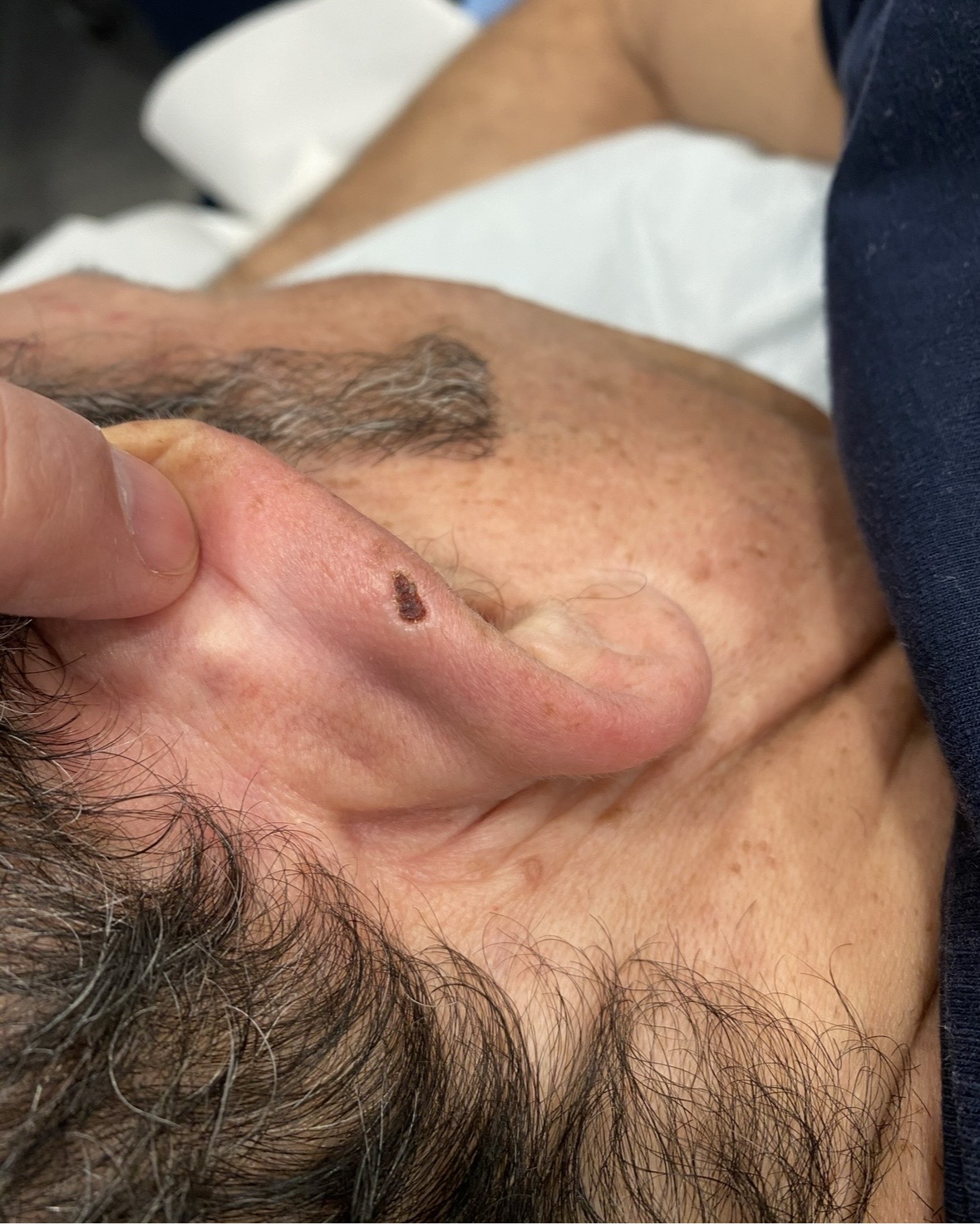
Pigmented Lesion of the Ear · Kare Skin Cancer Center · Dr. Sierro · Santa Monica Darkly pigmented, irregular lesion of the auricle evaluated with dermoscopy · Concerning features for melanoma or high-grade dysplastic nevus · Same-day biopsy performed during evaluation visit
Why the Ear Is High Risk
The ear is among the highest-risk anatomical locations for skin cancer — both melanoma and squamous cell carcinoma. The helical rim, anti-helix, and tragus receive intense cumulative UV exposure in a zone that most patients do not adequately protect with sunscreen. The ear is also an area that many patients and even some physicians fail to include in skin examinations. Any darkly pigmented, irregular, or evolving lesion on the ear requires prompt dermoscopy evaluation.
Dermoscopy Evaluation
This lesion demonstrates dermoscopic features that prompted same-day biopsy by Dr. Sierro — irregular pigment distribution, atypical pigment network at the lesion periphery, and color variation within the lesion consistent with concerning melanocytic activity. Dermoscopy evaluation of pigmented ear lesions is essential: the curved, convex anatomy of the auricle makes naked-eye assessment unreliable, and dermoscopy provides the subsurface structural analysis needed to make an accurate assessment.
Same-Day Action
Dr. Sierro performed an in-office excisional biopsy of this lesion under local anesthesia during the same visit at which it was identified — with complete excision and fine suture closure. The specimen was sent to dermatopathology for histological analysis. Same-day biopsy eliminates the dangerous gap between evaluation and action that allows thin melanomas to progress to invasive disease while patients wait for a return appointment.
Melanoma Warning Signs
The ABCDE Warning Signs of Melanoma
A Asymmetry One half of the mole does not mirror the other half in any direction
B Border Irregular, ragged, notched, blurred, or poorly defined edges
C Color Multiple colors — shades of brown, black, red, white, or blue within a single lesion
D Diameter Larger than 6mm — approximately the size of a pencil eraser — though melanoma can be smaller
E Evolving Any change in size, shape, color, or new symptoms — bleeding, itching, or crusting
Melanoma Staging — Why Stage at Diagnosis Determines Everything
Stage
Breslow Thickness
5-Year Survival
Treatment
I
<2mm, no ulceration, no nodes
98%
Wide local excision ± SLNB
II
>2mm or ulcerated, no nodes
65–93%
Wide local excision + SLNB
III
Any thickness, node involvement
40–78%
WLE + lymph node dissection + adjuvant
IV
Distant metastasis
<30%
Immunotherapy / targeted therapy
Dr. Sierro's Melanoma Management Protocol at the Kare Skin Cancer Center
Dermoscopy-Enhanced Full-Body Screening
Every mole evaluated at the Kare Skin Cancer Center receives dermoscopy — polarized light magnification that reveals the subsurface pigment network, vascular patterns, and structural features of melanoma invisible to the naked eye. Dr. Sierro applies evidence-based dermoscopy pattern analysis to assess each lesion for melanoma-specific structures: atypical pigment network, regression structures, blue-white veil, atypical vascular patterns, and peripheral globules. Dermoscopy improves melanoma detection sensitivity by up to 35% compared to visual examination alone.
Atypical Mole (Dysplastic Nevus) Management
Atypical moles (dysplastic nevi) are melanocytic nevi with abnormal architectural and cytological features that require individualized management based on histological grade. Mildly dysplastic nevi with clear margins may be observed with 6-monthly dermoscopy surveillance. Moderately and severely dysplastic nevi require re-excision with clear margins. Patients with multiple atypical moles — atypical mole syndrome — require surveillance every 6 months with total body photography for change detection. Dr. Sierro designs an individualized atypical mole protocol at each patient's first consultation.
Same-Day Biopsy
When Dr. Sierro identifies dermoscopic features concerning for melanoma, she performs an in-office biopsy under local anesthesia during the same visit — eliminating the dangerous delay between identification and tissue diagnosis. Excisional biopsy (complete removal with 1–3mm margins) is performed whenever feasible for small suspicious lesions — providing definitive diagnosis and initial treatment in a single procedure. Shave biopsy is avoided for suspected melanoma; proper depth assessment requires a full-thickness specimen.
Wide Local Excision & Staging Coordination
Confirmed melanoma triggers a comprehensive management protocol at the Kare Skin Cancer Center. Wide local excision with oncologically appropriate margins — 1cm for melanoma in situ, 1–2cm for invasive melanoma based on Breslow thickness — is performed by the plastic surgery team at Kare Plastic Surgery. Sentinel lymph node biopsy is coordinated with surgical oncology for tumors meeting SLNB criteria (Breslow thickness ≥0.8mm or ulceration). Medical oncology referral for immunotherapy or targeted therapy is arranged for Stage III–IV disease. No patient navigates a melanoma diagnosis without a clear management plan at Kare Plastic Surgery.
Total body photography for high-risk patients: Patients with atypical mole syndrome, personal history of melanoma, or family history of melanoma benefit from total body photography — a comprehensive photographic map of all moles that allows Dr. Sierro to detect new lesions or change in existing lesions at each 6-monthly surveillance visit. Subtle changes in mole size, shape, or color that are imperceptible without comparison photographs are clearly identified with this technique. Ask about total body photography at your Kare Skin Cancer Center consultation. Call (310) 998-5533.
"Early detection is key for melanoma survival."
— Kare Skin Cancer Center, Santa Monica
Frequently Asked Questions
What is the survival rate for melanoma caught early?
Stage I melanoma detected while thin and confined to skin has a 5-year survival rate exceeding 98%. Stage IV melanoma carries less than 30% 5-year survival. Early detection by Dr. Sierro at the Kare Skin Cancer Center in Santa Monica is the most effective melanoma mortality reduction strategy. Call (310) 998-5533.
What is an atypical mole and does it need to be removed?
Atypical moles (dysplastic nevi) are melanocytic nevi with abnormal features. Mildly dysplastic nevi with clear biopsy margins may be observed. Moderately and severely dysplastic nevi require re-excision with clear margins. Multiple atypical moles require 6-monthly surveillance. Dr. Sierro designs individualized protocols at Kare Plastic Surgery in Santa Monica. Call (310) 998-5533.
What does a melanoma look like on the ear?
Melanoma on the ear typically presents as a darkly pigmented, irregular lesion on the helical rim or auricle — an area of high cumulative UV exposure that many patients and physicians overlook. Any new, changing, or irregularly pigmented lesion on the ear requires same-day evaluation. Dr. Sierro performs same-day biopsy of suspicious ear lesions. Call (310) 998-5533.
Schedule Your Melanoma Screening
Appointments · Kare Skin Cancer Center Schedule a Melanoma Screening with Dr. Sierro →
Same-week melanoma screening, atypical mole evaluation, and biopsy appointments with Dr. Tiffany Sierro at Kare Plastic Surgery, Santa Monica. (310) 998-5533. Serving Los Angeles, Beverly Hills, and Pacific Palisades.
Authority Page · Kare Plastic Surgery Skin Cancer Treatment Los Angeles →
Comprehensive skin cancer diagnosis and treatment at the Kare Skin Cancer Center — melanoma, BCC, SCC, dermoscopy, biopsy, and wide local excision by Dr. Tiffany Sierro. (310) 998-5533.
Kare Skin Cancer Center · Kare Plastic Surgery · 804 7th Street, Santa Monica
Protect Yourself from Melanoma — Schedule a Screening with Dr. Sierro
Dr. Tiffany Sierro, board-certified dermatologist at the Kare Skin Cancer Center, provides expert melanoma screening, atypical mole management, and comprehensive skin cancer care throughout Santa Monica and Los Angeles — dermoscopy-enhanced evaluation, same-day biopsy, wide local excision, and coordinated staging. Same-week appointments available.