Kare Skin Cancer Biopsy Center · Kare Plastic Surgery · Santa Monica
Skin Cancer Biopsy Center Santa Monica
804 7th Street, Santa Monica, CA 90403 · (310) 998-5533
Same-day shave, punch, and excisional biopsy — skin cancer diagnosis, recurrence management, and post-treatment surveillance by board-certified dermatologist Dr. Tiffany Sierro at Kare Plastic Surgery
Call (310) 998-5533 Schedule a Biopsy Evaluation
BoardCertified Dermatologist
Same-DaySkin Biopsy
Skin Cancer Biopsy Center Santa Monica
A Suspicious Lesion Often Require a Biopsy
Dr. Tiffany Sierro, MD Board-Certified Dermatologist — Skin Cancer Biopsy Specialist Same-Day Biopsy · BCC · SCC · Melanoma · Recurrence · Surveillance · Kare Skin Cancer Biopsy Center, Santa Monica
The most dangerous gap in skin cancer care is not between finding a suspicious lesion and booking an appointment — it is between that appointment and the moment a biopsy is actually performed. Most dermatology practices require a dedicated biopsy appointment days or weeks after the initial evaluation. In that window, a lesion that presents as Stage I may progress. At the Kare Skin Cancer Biopsy Center, Dr. Tiffany Sierro, MD performs same-day biopsy of any suspicious lesion identified at the screening visit — closing that gap entirely.
Dr. Sierro's biopsy program begins with dermoscopy-enhanced evaluation of every concerning lesion. If dermoscopy identifies features consistent with basal cell carcinoma, squamous cell carcinoma, melanoma, or a high-grade atypical nevus, the biopsy is performed immediately under local anesthesia — shave, punch, or excisional, selected based on the lesion type and the diagnostic information required. The specimen proceeds to a board-certified dermatopathologist that same day. Results are reviewed with patients promptly, and the treatment plan is communicated with the same urgency as the biopsy.
Skin cancer recurrence requires immediate evaluation. Any new growth, ulceration, redness, or firmness at or near the site of a previously treated skin cancer is a recurrence until proven otherwise. Do not wait for a routine annual appointment. Call Dr. Sierro at the Kare Skin Cancer Biopsy Center at (310) 998-5533 for a same-week recurrence evaluation.
Kare Skin Cancer Biopsy Center · Santa Monica · (310) 998-5533
Suspicious lesion, prior skin cancer, or due for a screening? Schedule your same-day biopsy evaluation with Dr. Sierro today.
Clinical Documentation — Kare Skin Cancer Biopsy Center · Dr. Tiffany Sierro
Skin Cancer Recurrence — Evaluated and Biopsied at the Kare Skin Cancer Center
Actual patient at Kare Plastic Surgery · Recurrent skin cancer lesion at prior treatment site · Dermoscopy evaluation and same-day excisional biopsy by Dr. Tiffany Sierro
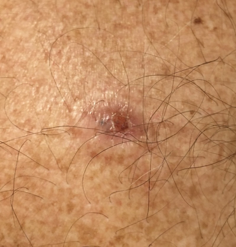
Skin Cancer Recurrence · Kare Skin Cancer Center · Dr. Sierro · Santa Monica Recurrent pigmented erosive lesion at prior skin cancer treatment site · Background photodamaged skin · Dermoscopy evaluation performed · Same-day excisional biopsy for histological diagnosis
Recognizing Recurrence
This patient presents with a recurrent pigmented, erosive lesion at a site of prior skin cancer treatment — a presentation that requires immediate dermoscopy evaluation and same-day biopsy. The lesion demonstrates irregular pigmentation, surface erosion, and surrounding erythema on a background of diffuse solar damage consistent with a prior BCC or SCC treatment site. Any change at a previously treated skin cancer site is a recurrence until the biopsy says otherwise.
Why Recurrences Are More Complex
Recurrent skin cancers are more challenging to treat than primary lesions — scar tissue from the prior treatment alters the tissue planes, may conceal the extent of tumor invasion, and creates a less predictable surgical field. Recurrent BCC in particular is associated with a higher rate of incomplete excision and subsequent re-recurrence than primary BCC. Dr. Sierro evaluates every recurrent skin cancer with dermoscopy and selects the management strategy — re-excision, Mohs coordination, or systemic therapy — based on the histological findings and clinical context.
Dr. Sierro's Response
Same-day excisional biopsy was performed under local anesthesia during this patient's evaluation visit. The specimen was immediately sent to dermatopathology for margin assessment and subtype characterization. For recurrent lesions, histological subtype is critical: recurrent morpheaform BCC and recurrent perineural SCC carry substantially higher re-recurrence risk and require referral for Mohs surgery rather than standard excision. Dr. Sierro communicates results within days and coordinates the definitive treatment plan.
Biopsy Techniques
Skin Biopsy Options at Kare Skin Cancer Biopsy Center
Shave Biopsy
A thin blade is used to remove a superficial horizontal tissue sample — appropriate for raised, dome-shaped, or exophytic lesions including seborrheic keratoses, superficial basal cell carcinomas, dermatofibromas, and viral warts. Shave biopsy is quick, requires no sutures, and heals well on most body sites. It is not appropriate for suspected melanoma — where a full-thickness specimen with depth information is required — or for pigmented lesions where Breslow thickness measurement would be needed for staging. Dr. Sierro uses shave biopsy when the clinical and dermoscopic features indicate a benign or superficial diagnosis and when complete depth assessment is not required.
Punch Biopsy
A circular cutting tool (2–6mm diameter) is rotated through the full thickness of the skin to obtain a cylindrical tissue sample including epidermis, dermis, and upper subcutaneous fat. Punch biopsy is the most versatile technique — appropriate for pigmented flat lesions, inflammatory conditions, suspected BCC and SCC, and diagnostic sampling of larger lesions where excisional biopsy is not practical. A single suture closes the defect. Punch biopsy provides full-thickness tissue for accurate histological assessment of lesion depth and dermal involvement.
Excisional Biopsy
The entire lesion is removed with a margin of surrounding normal skin using an elliptical excision under local anesthesia — providing both diagnosis and treatment in a single procedure for smaller lesions. Excisional biopsy is the preferred technique for suspected melanoma (complete depth measurement of the entire lesion required for staging), atypical nevi, and small skin cancers where diagnostic and therapeutic goals align. The specimen is oriented with suture marks for the pathologist to assess margin status. Layered suture closure produces a fine, linear scar.
Incisional Biopsy
A partial sample of a larger lesion is obtained through elliptical excision — used when the lesion is too large for complete excisional biopsy in the office setting, or when the clinical diagnosis requires tissue confirmation before planning a larger definitive excision. Incisional biopsy is appropriate for large suspected BCCs, large actinic keratoses, suspected Merkel cell carcinoma, and any lesion where initial diagnostic sampling informs the subsequent treatment decision. Dr. Sierro selects the most appropriate biopsy approach at each patient's evaluation.
Skin Cancer Recurrence — Who Is at Risk and What to Watch For
Prior BCC or SCC
Patients with a history of basal cell carcinoma or squamous cell carcinoma have a 40–50% risk of developing a new skin cancer within 5 years. Regular 6-monthly surveillance by Dr. Sierro allows new lesions to be identified and biopsied before they require complex treatment.
Aggressive Histological Subtypes
Morpheaform BCC, perineural SCC, and invasive SCC carry significantly higher recurrence rates than common nodular BCC. Prior treatment of these aggressive subtypes warrants more intensive surveillance and a lower threshold for biopsy of any new change at the treatment site.
Incompletely Excised Lesions
When prior skin cancer pathology reports a positive or close margin, local recurrence risk is substantially elevated. Dr. Sierro reviews prior pathology reports at every new patient consultation and designs a surveillance protocol based on the prior excision margin status and tumor subtype.
Immunosuppressed Patients
Organ transplant recipients, patients on chronic immunosuppressive therapy, and HIV-positive patients have dramatically elevated skin cancer risk — particularly for SCC, which can be aggressive and multiply recurrent in the immunosuppressed setting. Intensive 3-monthly surveillance is standard for this population at Kare Plastic Surgery.
Melanoma History
Patients with a personal history of melanoma require lifelong surveillance — 3-monthly for the first 2 years, 6-monthly for years 3–5, then annually. Any new pigmented lesion or change at the primary excision site or lymph node basin warrants immediate evaluation by Dr. Sierro and same-day biopsy when clinically indicated.
Field Cancerization
Extensive sun-damaged skin with multiple actinic keratoses represents a field of UV-mutated keratinocytes at elevated risk for SCC development throughout the affected zone. Dr. Sierro identifies and treats field cancerization with Efudex, imiquimod, or PDT — and biopsies any area within the field that fails to respond or develops an indurated or erosive lesion.
Post-treatment surveillance program at Kare Plastic Surgery: Every patient treated for a skin cancer at the Kare Skin Cancer Biopsy Center is enrolled in a post-treatment surveillance schedule — 6-monthly dermoscopy screening for BCC and SCC patients, 3-monthly screening for melanoma patients during the first 2 years. Surveillance appointments include full-body dermoscopy evaluation, dermoscopy documentation of all existing nevi, and same-day biopsy of any new or changing lesion. Call (310) 998-5533 to establish your surveillance program with Dr. Sierro.
Frequently Asked Questions
What happens during a skin biopsy at Kare Plastic Surgery in Santa Monica?
Dr. Sierro performs an in-office biopsy under local anesthesia — selecting shave, punch, or excisional technique based on the lesion. The procedure takes 10–20 minutes. The specimen is sent to dermatopathology and results are returned within 7–10 business days. Dr. Sierro reviews all results with patients and determines the next steps. Call (310) 998-5533.
Can skin cancer come back after treatment?
Yes. Patients with prior BCC or SCC have a 40–50% risk of a new skin cancer within 5 years. Recurrence at the treatment site, new primaries, and — for high-risk SCC and melanoma — regional or distant recurrence are all recognized risks. Regular post-treatment surveillance with Dr. Sierro at Kare Plastic Surgery is essential. Call (310) 998-5533.
What is the difference between a shave biopsy, punch biopsy, and excisional biopsy?
Shave biopsy removes a superficial sample — used for raised lesions. Punch biopsy removes a full-thickness cylinder — for flat lesions and BCC/SCC diagnosis. Excisional biopsy removes the entire lesion — preferred for suspected melanoma. Dr. Sierro selects the appropriate technique at each evaluation. Call (310) 998-5533.
Schedule Your Biopsy Evaluation
Kare Skin Cancer Biopsy Center · Kare Plastic Surgery · 804 7th Street, Santa Monica
Get Your Skin Cancer Biopsy Done Today with Dr. Sierro
Dr. Tiffany Sierro, board-certified dermatologist at the Kare Skin Cancer Biopsy Center, provides same-day biopsy and comprehensive skin cancer recurrence management throughout Santa Monica and Los Angeles — dermoscopy evaluation, same-day tissue sampling, dermatopathology coordination, and individualized post-treatment surveillance programs. Same-week appointments available.