Kare Plastic Surgery · Santa Monica · Los Angeles
Biopsy and Ultrasound Guided Mapping of Facial Fillers Los Angeles
804 7th Street, Santa Monica, CA 90403 · (310) 998-5533
Dr. Raffy Karamanoukian at Kare Plastic Surgery uses high-frequency ultrasound mapping and tissue biopsy to identify filler type, location, and complications before removal throughout Los Angeles. The most precise filler complication workup available.
Schedule a Consultation Filler Removal Overview
Raffy Karamanoukian MD FACSDual Board-Certified Surgeon
Ultrasound Guided Filler RemovalReal-Time Precision Mapping
Tissue BiopsyFiller Type Confirmation
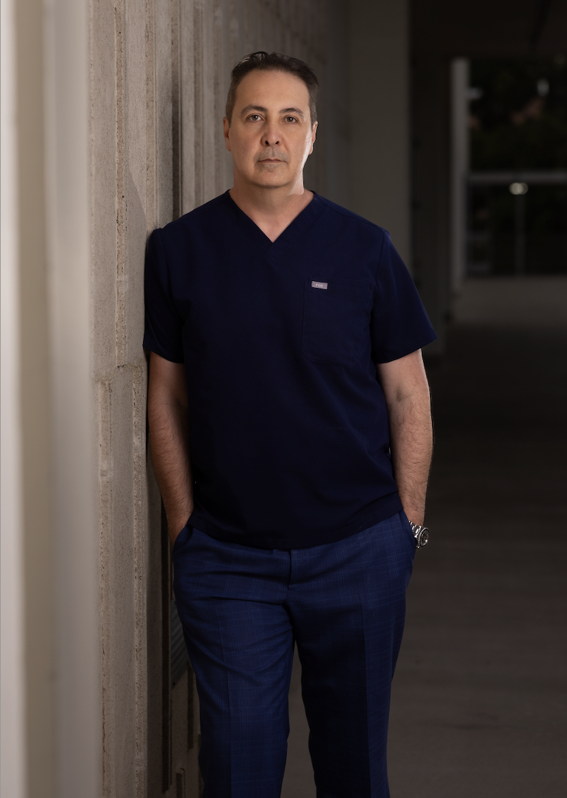
Dr. Raffy Karamanoukian, MD FACS Dual Board-Certified Plastic Surgeon · Filler Complication Specialist Kare Plastic Surgery · 804 7th Street, Santa Monica, CA 90403 · (310) 998-5533
Filler Ultrasound Mapping and Biopsy · Los Angeles
Precision Filler Diagnosis: Ultrasound Mapping | Tissue Biopsy
When a filler complication occurs, the most important first step is knowing exactly what type of material was injected, where it is, how far it has spread, and what the surrounding tissue looks like. Without this information, any attempt at removal risks being incomplete, misdirected, or unsafe.
Dr. Raffy Karamanoukian at Kare Plastic Surgery uses two powerful diagnostic tools before every filler complication treatment: high-frequency ultrasound mapping to visualize filler deposits in real time, and tissue biopsy to confirm the filler type under the microscope when the material is unknown or a granuloma or nodule has formed. Together these two tools give Dr. Karamanoukian a complete picture of the problem before any treatment begins. Learn more on our ultrasound guided filler removal page. Call (310) 998-5533.
Kare Plastic Surgery · Santa Monica, Los Angeles
Schedule A Filler Complication Consultation Dr. Karamanoukian
Ultrasound Mapping and Tissue Biopsy at Kare Plastic Surgery

Ultrasound Filler Mapping Filler ultrasound showing subcutaneous filler deposit during mapping by Dr. Karamanoukian. Anechoic and hyperechoic patterns distinguish filler type before removal planning.
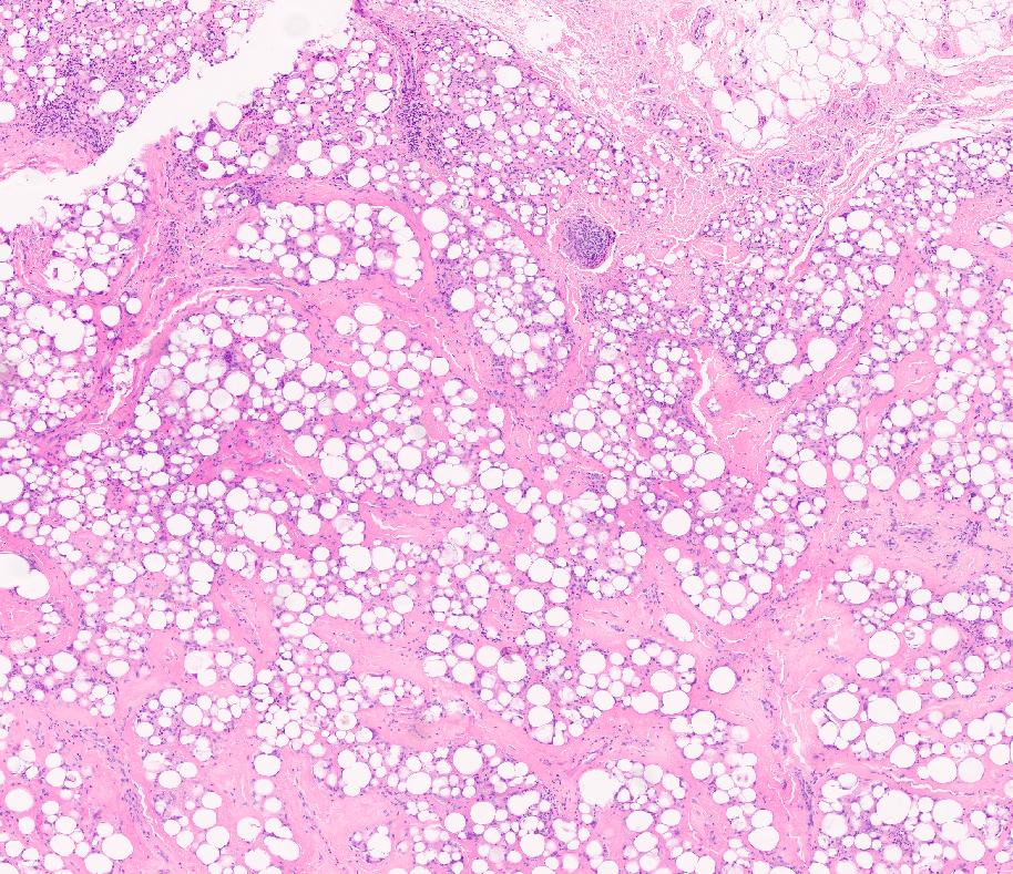
Tissue Biopsy · Filler Histopathology Hematoxylin and eosin histopathology slide from a filler tissue biopsy showing the composition of a filler-affected tissue sample. Used to confirm filler type and granuloma formation before treatment at Kare Plastic Surgery.
Ultrasound Mapping and Biopsy: How Each Tool Is Used
High-Frequency Ultrasound Mapping
Ultrasound reveals filler location, depth, spread, and proximity to blood vessels in real time without any incision. Each filler type produces a distinct pattern: HA fillers appear as anechoic (dark) pockets, PMMA creates bright echogenic deposits with comet-tail artifacts, and liquid silicone produces a snowstorm reverberation pattern. Ultrasound also identifies vascular structures that must be avoided during removal and can detect oil cysts from fat grafting that are present alongside filler. This mapping information guides every subsequent treatment decision.
Tissue Biopsy
When the injected material is unknown, or when a nodule or granuloma has formed, a tissue sample is taken and sent to pathology for hematoxylin and eosin staining and special stains. Biopsy confirms whether the material is hyaluronic acid, PMMA, silicone, or another foreign body, and whether the tissue shows granulomatous inflammation, foreign body giant cells, or fibrosis. This information determines whether hyaluronidase can be used, whether surgery is required, and the expected degree of difficulty of removal. Biopsy also rules out infection and malignancy in any uncertain eyelid or facial nodule.
Filler Types Identified at Kare Plastic Surgery
Hyaluronic Acid
Anechoic on ultrasound. Dissolved with hyaluronidase. Biopsy shows mucin deposits with Alcian blue stain.
PMMA (Bellafill)
Echogenic deposits with comet-tail artifact on ultrasound. Not dissolvable. Requires surgical excision.
Silicone and Biopolymers
Snowstorm ultrasound pattern. Biopsy shows foreign body giant cells. Requires open surgical removal.
Kare Plastic Surgery is one of the only centers in Los Angeles offering both ultrasound mapping and formal tissue biopsy as part of a complete filler complication workup before any removal procedure is performed. Most injectors do not have access to high-frequency ultrasound or pathology processing. Dr. Karamanoukian's dual board certification in plastic surgery provides the surgical and diagnostic depth to manage complex filler complications safely. Call (310) 998-5533.
Frequently Asked Questions
Why is ultrasound used to map facial fillers before removal?
Ultrasound may show filler location, depth, and spread and identifies blood vessels nearby. Different fillers produce distinct ultrasound patterns that help identify the material before removal. Dr. Karamanoukian at Kare Plastic Surgery uses ultrasound mapping before filler removal procedure. Call (310) 998-5533.
When is a tissue biopsy needed for a filler complication?
Biopsy is needed when the filler type is unknown, when a nodule or granuloma has formed and needs histological confirmation, or to rule out infection or malignancy. Pathology stains confirm whether HA, PMMA, silicone, or another foreign body is present, which determines the correct removal approach.
Can all fillers be dissolved with hyaluronidase?
Hyaluronic acid fillers respond to hyaluronidase. PMMA, silicone, and biopolymers require surgical removal. Biopsy and ultrasound mapping confirm which approach is correct before any treatment begins. Call our office for a filler complication evaluation at Kare Plastic Surgery in Los Angeles.
Schedule Your Filler Complication Evaluation
Kare Plastic Surgery · Los Angeles & Santa Monica · 804 7th Street
Diagnose Your Filler Complication with Dr. Karamanoukian
Dr. Raffy Karamanoukian, dual board-certified plastic surgeon at Kare Plastic Surgery, offers ultrasound guided filler mapping and tissue biopsy for complete filler complication diagnosis throughout Los Angeles. Same-week appointments available.